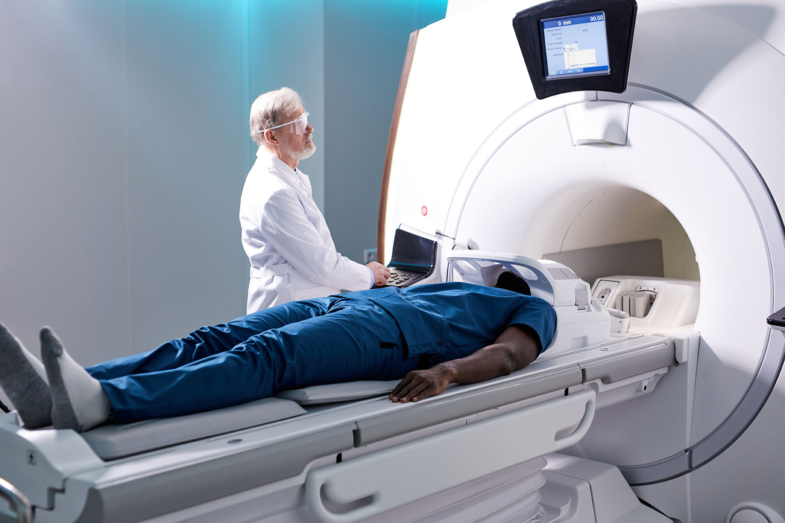

Radiology is the use of ionizing radiation to create images of the body for diagnostic and therapeutic purposes. Ionizing radiation is radiation that has enough energy to knock electrons out of atoms. This can damage cells and tissues, but it can also be used to create images of the body.
The most common type of radiation used in radiology is X-rays. X-rays are a form of ionizing radiation that can penetrate the body and create images of bones, organs, and other structures. Other types of radiation used in radiology include:
Radiology is a safe and effective way to diagnose and treat medical conditions. However, it is important to remember that radiation can be harmful to the body. Therefore, it is important to use radiation only when necessary and to follow the guidelines for safe radiation exposure.
The radiologist is a doctor who specialises in radiology.

Noun: the branch of medical science concerned with the use of radiant energy (such as X-rays) in the diagnosis and treatment of disease.
Adjective: of or relating to radiology.
The word "radiology" is a combination of the words "radio-" and "logy".
The word "radio-" comes from the Latin word "radius", which means "ray".
The suffix "-logy" comes from the Greek word "logos", which means "study".
The first recorded use of the word "radiology" was in 1896.
What does a Radiologist do?
Question:
Define radiology and explain how X-rays are used in medical imaging. Provide an example of a medical condition where radiology plays a crucial role in diagnosis.
Answer:
Radiology is a branch of medical science that involves the use of various imaging techniques to visualize the internal structures of the human body. X-rays, a common tool in radiology, are a form of electromagnetic radiation with higher energy than visible light. They are used for medical imaging to create detailed images of bones, tissues, and organs.
In medical imaging, X-rays work by passing through the body and being absorbed by different tissues to varying degrees. Dense tissues, like bones, absorb more X-rays, appearing as white areas on the X-ray image. Less dense tissues, like muscles and organs, allow more X-rays to pass through, resulting in darker areas on the image.
For instance, consider a patient who experiences persistent chest pain. To diagnose the cause, a doctor may request a chest X-ray. This non-invasive imaging technique can reveal if there are any fractures, infections, or abnormalities in the lungs, heart, or ribs. The resulting X-ray image can provide valuable information that aids in determining the appropriate course of treatment.
Radiology is integral to modern medicine, offering insights into the body's internal structures without invasive procedures. X-rays, among other imaging methods, allow medical professionals to diagnose conditions accurately, plan interventions, and monitor progress, thereby contributing significantly to patient care and treatment decisions.